Figure 3.

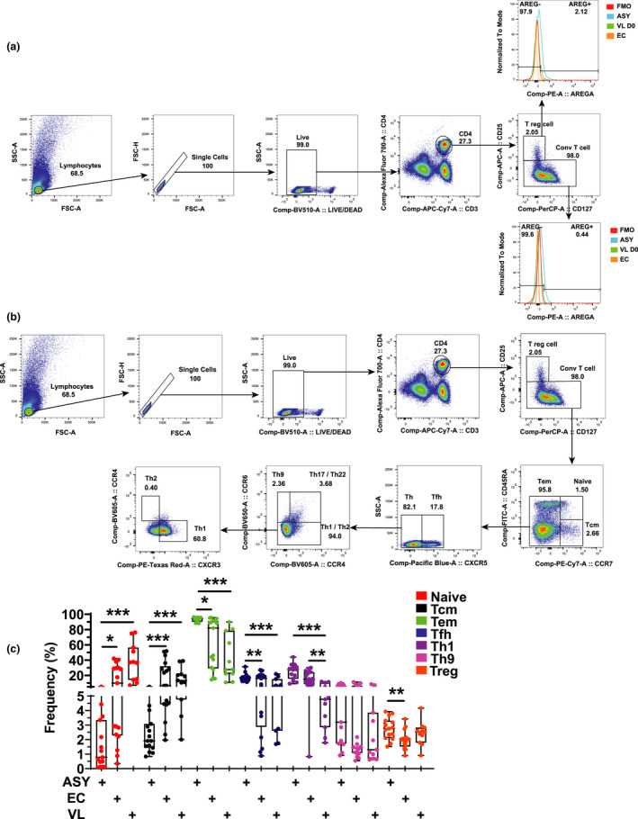
Changes in CD4+ T‐cell subset frequencies and expression of AREG. CD4+ T cells were identified by CD3ε and CD4 expression prior to being divided into regulatory T (Treg) cells and conventional T cells, based on CD25 and CD127 expression, before assessing AREG expression (a). CD4+ T cells were divided into T helper cell subsets based on chemokine receptor expression (b) and frequencies measured in peripheral blood from asymptomatic (ASY; n = 15) individuals, endemic controls (EC; n = 17) visceral leishmaniasis patients (VL; n = 11). The box shows the extent of the lower and upper quartiles plus the median, while the whiskers indicate the minimum and maximum data points (c). *P < 0.05, **P < 0.01 and ***P < 0.001. Significance was assessed by the Kruskal–Wallis test.
