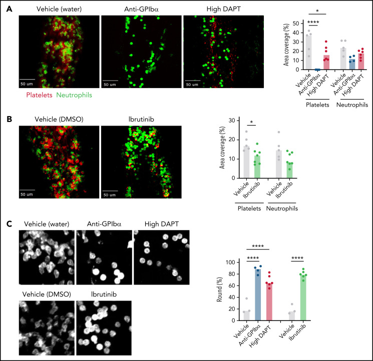
Figure 6.
Both platelet GPCR and ITAM receptor signaling contribute to PNA formation on the endothelium during VT initiation. WT mice were treated with anti-GPIbα antibodies to deplete circulating platelets, or with high DAPT (A), or ibrutinib (B) and then subjected to saphenous vein ligation. Spinning disk confocal intravital microscopy was used to visualize platelet (anti-GPIX antibody, red) and neutrophil (anti-Ly6G antibody, green) adhesion to the saphenous vein wall. Representative still frames from the videos taken after 2 hours of flow restriction and quantification of the area covered by platelets or neutrophils are shown. Dots represent individual mice, bars indicate medians. (C) Left panel: higher magnification images illustrating altered morphology of adherent neutrophils in mice treated with anti-GPIbα antibodies, high DAPT, or ibrutinib. Right panel: Quantification of percent round cells in selected fields of view. Dots represent individual mice, bars indicate medians. *P < .05, ****P < .0001.

