Figure 1.
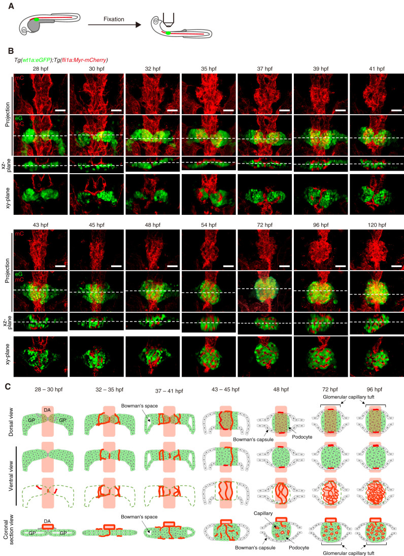
Morphogenetic processes of glomerular capillary formation in zebrafish pronephros. (A) Schematic illustration showing a sample preparation procedure and imaging of the zebrafish pronephros. After removal of the yolk sac from the fixed embryos and larvae, they were mounted ventral side up in 1% low-melting agarose and imaged as described in the Materials and Methods section. (B) Confocal z-projection and single-plane images of the zebrafish pronephros in Tg (wt1a:eGFP);Tg(fli1a:Myr-mCherry) embryos and larvae at the stage indicated at the top of each column. Ventral views; anterior to the top. The first and second rows show z-projection images of fli1a:Myr-mCherry (mC; red) and the merged images of wt1a:eGFP (eG; green) and fli1a:Myr-mCherry (mC; red), respectively. The third row is cross-sectional single-plane images (xz-plane) of the areas indicated by dotted lines on the images in the second row. The bottom row shows the xy-single slice images of the areas indicated by dotted lines on the xz-plane images. Scar bars: 20 μm. (C) Schematic drawings of the glomerular structures in the zebrafish pronephros at the stage indicated at the top of each column. Green indicates wt1a:eGFP-positive glomerular primordia and podocytes, whereas red shows fli1a:Myr-mCherry-positive blood vessels. The first row is the dorsal view images of the glomeruli, whereas the second and third rows are the ventral view images that correspond to the z-projection images in (B). In the third row, the wt1a:eGFP-positive glomerular primordia and podocytes are indicated by dotted green lines. The bottom row shows the coronal section views of the glomeruli corresponding to the cross-sectional single-plane images in (B). GP, glomerular primordia; DA, dorsal aorta.

