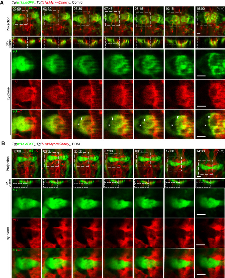
Figure 5.
Glomerular morphogenesis in the presence and absence of blood flow. Confocal fluorescence images of pronephric glomeruli in 33 hpf Tg(wt1a:eGFP);Tg(fli1a:Myr-mCherry) embryos in the absence (A) or presence (B) of BDM and the corresponding subsequent time-lapse images, obtained at the indicated time points (h:min). Merged images of z-projections of wt1a:eGFP (eG; green) and fli1a:Myr-mCherry (mC; red) are shown in the top row. The second row shows the cross-sectional single-plane images (xz-plane) of the areas indicated by dotted lines on the images in the top row. The boxed areas on the images in the first row are enlarged as xy-plane images obtained at the z-levels indicated by the dotted lines on the xz-plane images in the three rows from the bottom, in which wt1a:eGFP images, fli1a:Myr-mCherry images, and their merged images are shown in the third, fourth, and fifth rows, respectively. Arrowheads, podocytes or podocyte progenitors moving toward the midline; asterisks, Bowman’s space. Scale bars: 20 μm.

