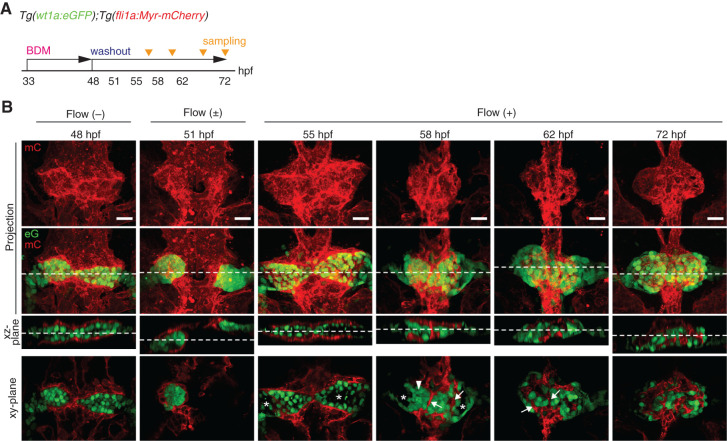
Figure 6.
Potential roles of blood filtration in glomerular remodeling and capillary formation. (A) Schematic diagram showing the experimental protocol for investigating the effects of stopping and subsequently restarting blood flow on glomerular morphogenesis and capillary formation. (B) The z-projection and single-plane images of the glomerular primordia and glomeruli in Tg(wt1a:eGFP);Tg(fli1a:Myr-mCherry) embryos are shown as in Figure 1B. The 48 hpf embryos treated with BDM from 33 hpf exhibited wt1a:eGFP-positive bilateral glomerular primordia covered by the fli1a:Myr-mCherry-labeled endothelial cells sprouted from the dorsal aorta (leftmost column). After termination of BDM treatment from 48 hpf, blood circulation was detected in the embryos at 51 hpf (second column from the left). Subsequently, structural changes in the vascular sheets and the enveloping glomerular primordia associated with the onset of blood flow were analyzed at 55, 58, 62, and 72 hpf (four columns from the right). Arrowheads, blood vessels that were derived from the vascular sheets enveloping the glomerular primordia; asterisks, inner cavity formed inside the glomerular primordia or Bowman’s space. Scale bars: 20 μm.

