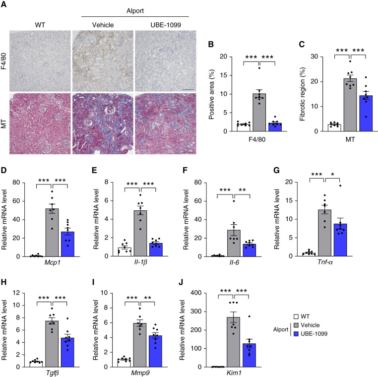
Figure 4.
UBE-1099 suppressed renal tissue inflammation and fibrosis in an Alport mouse model. (A) Renal sections of 22-week-old WT and Alport mice were analyzed using F4/80 immunohistochemistry and Masson’s trichrome (MT) staining. Representative images are shown. Scale bars: 200 μm. (B) and (C) F4/80-positive area and fibrotic region were evaluated based on the F4/80-stained section and MT-stained section, respectively, using Bio-Revo imaging and analysis software. (D–J) Total RNA was isolated from renal tissues of 22-week-old WT and Alport mice. The level of the indicated mRNA was measured and normalized to the level of Gapdh mRNA (internal control). Data are presented as the mean±SEM (n=7–8 per group). P values were assessed by Dunnett’s test. *P<0.05, **P<0.01, ***P<0.001.

