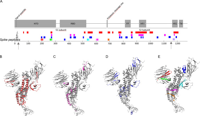
Figure 3.
NE cleavage sites within full-length S-protein. Arrows indicate limits of the cryo-EM structure 27–1146. (A) Peptides identified by LC-MS/MS (Table S1) in correlation to the amyloidogenic Spike peptides (cf. Figure 1 and Table 1) and the S-protein domain structure: after (i) 1 min (red), (ii) 1 min and still persistent at 6 h (magenta), (iii) only present after 6 h incubation (blue). (B–E) Same color code for cleaved peptide groups (i)–(iii) and spike peptides mapped onto the protomer cryo-EM S-protein structure PDB 6VXX.1

