Fig. 3.
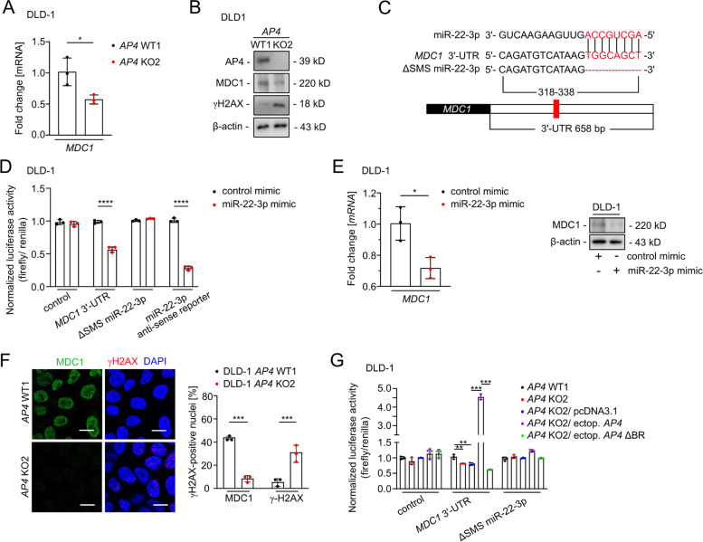
AP4-mediated repression of miR-22-3p contributes to increased MDC1 levels. A qPCR analysis of MDC1 expression. B Western blot analysis. C Scheme of the miR-22-3p seed, the seed-matching sequences and its deletion in the 3′-UTR in the MDC1 mRNA. The seed and seed-matching sequences are highlighted in red. D Dual-luciferase assay was conducted 48 h after DLD-1 cells were transfected with miR-22-3p mimic and human MDC1 3′-UTR reporter vector. The miR-22-3p anti-sense reporter served as a positive control. E qPCR (left panel) and Western blot analysis (right panel) 48 h after transfection. F MDC1 foci detection in untreated cells. Quantification of 3 fields with 120 cells in total. Scale bars: 20 μm. G Dual-luciferase assay 48 h after transfection. In panels A, D-G, the mean + SD (n = 3) is provided with *: p < 0.05, **: p < 0.01, ***: p < 0.001, ****: p < 0.0001

