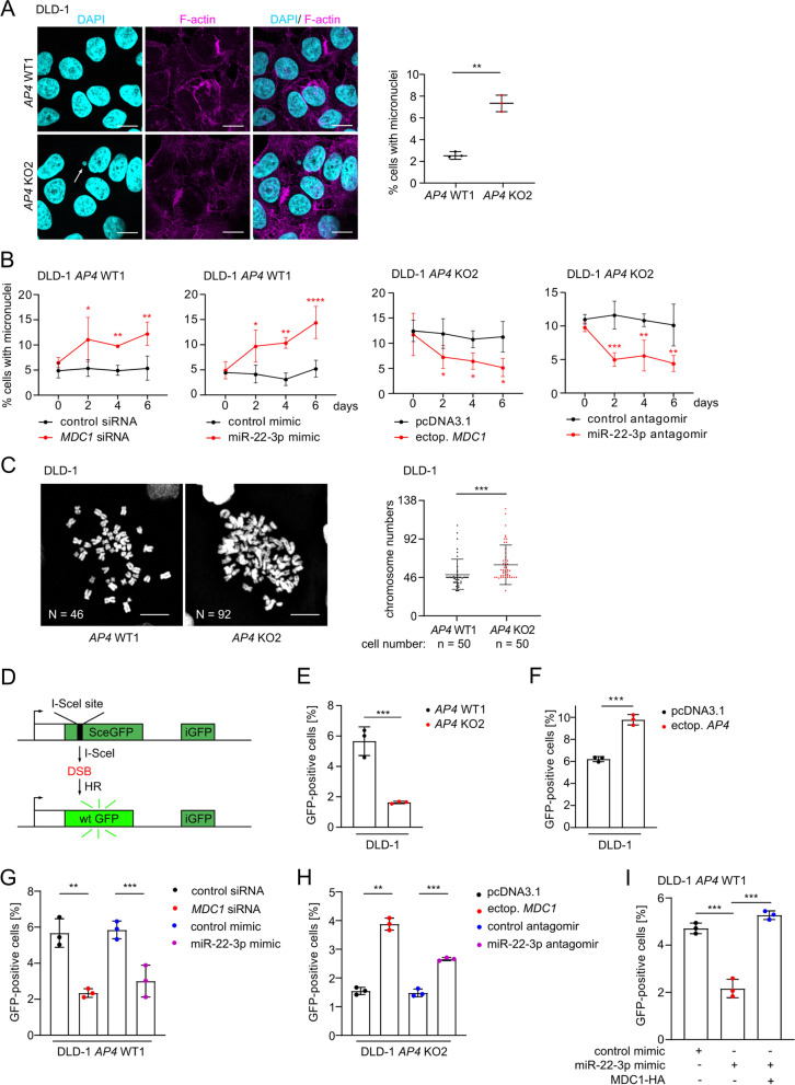
Fig. 8.
MDC1 mediates effects of AP4 on chromosomal instability. A Examples and quantification of micronuclei after DAPI staining. Three fields of 120 cells in total were evaluated. Scale bars: 20 μm. B Kinetic evaluation of micronucleus formation 48 h after transfection with the indicated oligonucleotides or vector. C Representative images of mitotic chromosome spreads. Quantification of 50 spreads per genotype. Scale bars: 20 μm. D Scheme illustrating the assay used for the fluorescence based measurement of HR-mediated DSB repair. E The indicated cells were co-transfected with pDR-GFP and pCBAScel plasmids. A pcDNA-mCherry plasmid was co-transfected as a control of transfection efficiency. The percentage of cells expressing GFP was measured by flow cytometry. F-I The percentage of the indicated cells expressing GFP was measured by flow cytometry 72 h after transfection of the indicated plasmids or oligonucleotides. In A-B and E-I the mean + SD (n = 3) and in C the mean + SD (n = 50) are provided with *: p < 0.05, **: p < 0.01, ***: p < 0.001, ****: p < 0.0001

