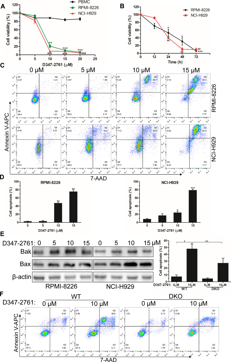
Fig. 2.
D347-2761 induced cytotoxicity in MM cells. A RPMI-8226 and NCI-H929 cells were treated by different dose of D347-2761 (5 μM, 10 μM, 15 μM and 20 μM) for 48 h and cell viabilities were measured using CCK8 kit. B Indicated cells treated by 10 μM D347-2761 at different time were dealt with CCK8 and cell survival rates were measured. C, D Flow cytometry analysis of apoptotic cell rates in RPMI-8226 and NCI-H929 cells by D347-2761 treatment (5 μM, 10 μM and 15 μM) for 48 h. E Western blot assay of Bak and Bax apoptotic markers in RPMI-8226 and NCI-H929 cells treated by D347-2761. β-actin was used to be internal control. F RPMI-8226-WT and –DKO cells were treated by 10 μM D347-2761 for 48 h and cell apoptosis was detected using Annexin V-APC/7-AAD apoptosis detection kit. Error bars: mean ± SD from at least three independent experiments. #, *P < 0.05, ##, **P < 0.01, ###, ***P < 0.001

