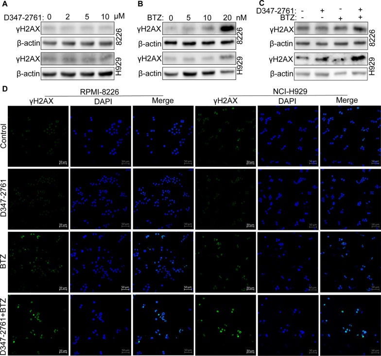
Fig. 3.
D347-2761 synergized with BTZ to promote DNA damage. A RPMI-8226 and NCI-H929 cells were treated by different dose of D347-2761 (2 μM, 5 μM and 10 μM) and western blot analysis of γH2AX expression. B Western bolt showing the expression of γH2AX in RPMI-8226 and NCI-H929 cells via different concentration of BTZ (5 nM, 10 nM and 20 nM). C RPMI-8226 and NCI-H929 cells were treated by 10 μM D347-2761 and/or 10 nM BTZ and western blot analysis of γH2AX expression. β-actin was used to be internal control. D Immunofluorescence assay of γH2AX expression level in indicated cells via treatment with 10 μM D347-2761 and/or 10 nM BTZ. The nucleus was stained by 1 μg/ml DAPI. Scale bars: 50 μm

