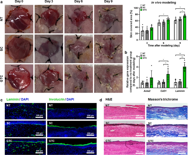
Fig. 7.
Improved in vivo wound healing in green OLED-treated hADSCs. a Representative photographs of skin wounds at 0, 3, 6, and 9 days after skin wound modeling and various treatments with wound coverage ratio quantification (n = 5). Mice were divided into three groups: no treatment (NT) served as control, hADSCs (SC), and green OLED-treated hADSCs (GTC) groups. b Gene expression levels of Acta2, Cd31, and Laminin in skin wound regions on day 9 as evaluated by qRT-PCR (n = 5). c Laminin (blue: nuclei, green: laminin, scale bar = 100 µm) and involucrin staining (blue: nuclei, green: involucrin, scale bar = 250 µm) of vertical sections of skin wound regions of each group on day 9. d H&E (scale bar = 200 µm) and Masson’s trichrome staining (scale bar = 200 µm) of vertical sections of skin wound regions of each group on day 9. *p < 0.05; **p < 0.001 compared to other groups with unpaired t test

