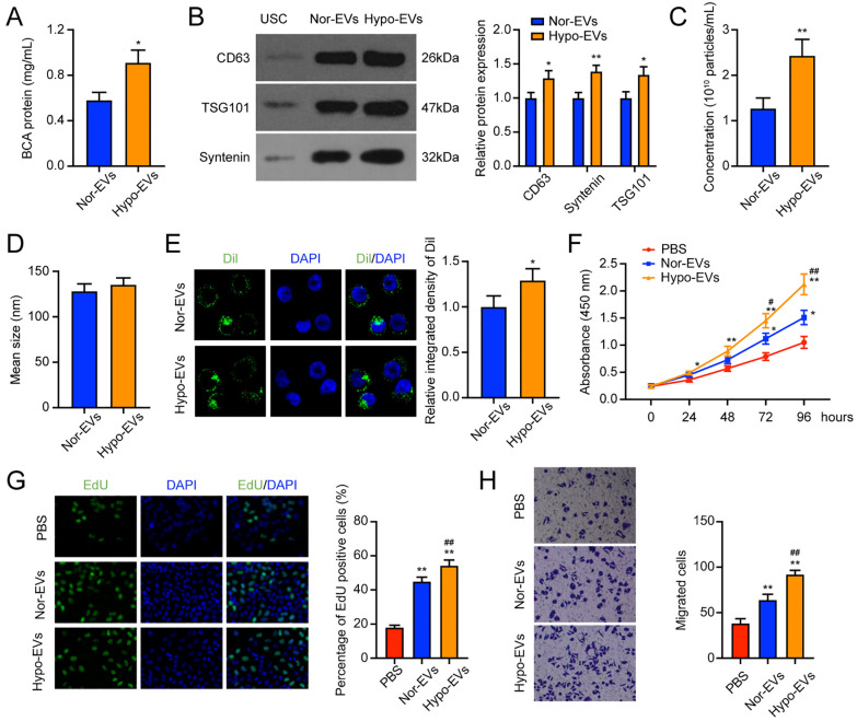
Figure 3.
Hypo-EVs enhanced the promoting effect of USC-EVs on chondrocytes. (A) BCA method to determine the protein concentration of the nor-EVs and hypo-EVs. (B) Western blot detection of EV surface biosignatures CD63, Syntenin, and TSG101. (C-D) The concentration (C) and mean size (D) of nor-EVs and hypo-EVs. (E) Dil-labeled EVs in chondrocytes. (F) CCK-8 assay detection of the cell viability of chondrocytes. (G) EdU staining of the cell proliferation of chondrocytes. (H) Transwell assay was applied to detect the migration ability of chondrocytes. EVs = extracellular vehicles; USCs = urine-derived stem cells; BCA = bicinchoninic acid; EdU = 5-Ethynyl-2’-Deoxyuridine; DAPI = 4’,-6-diamidino-2-phenylindole; PBS = phosphate-buffered saline; CCK-8 = cell counting kit-8. *P < 0.05, **P < 0.01, compared with the PBS group; #P < 0.05, ##P < 0.01, compared with the Nor-EVs group.

