Figure 1.
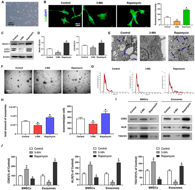
Autophagy regulates exosome release by MSCs. (A) BMSCs cultured in medium; scale bars = 100 μm. (B) Immunofluorescence staining depicting LC3+ cells (green) and quantitative analysis of intensity; scale bars = 20 μm. (C, D) Western blot analysis of Beclin1, LC3I, LC3II, and β-actin expressions in BMSCs. (E) TEM depicting autophagosomes (arrows); scale bars = 1 μm. (F) TEM observation of exosomes from BMSCs; scale bars = 200 nm. (G, H) NTA of the size distribution and amount of exosomes from BMSCs. (I, J) Western blot analysis of CD63, ALIX and TSG101 expressions in BMSCs and exosomes. MSCs = mesenchymal stem cells; BMSCs = bone marrow-derived MSCs; TEM = transmission electron microscopy; NTA = nanoparticle tracking analysis; DAPI: diamidine phenylindole; 3-MA = 3-methyladenine. The values are the mean ± SD; n = 3, *P < .05.

