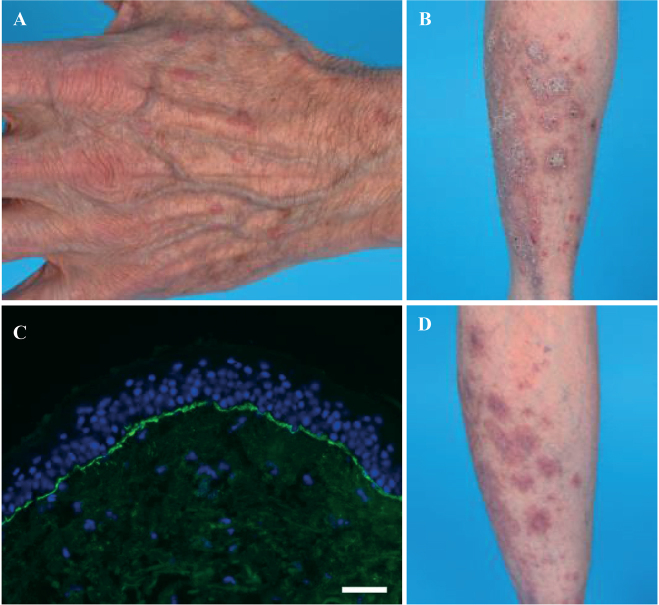
Fig. 1.
(A) Livid erythematous polygonal papules on the dorsal side of the hand. (B) Hyperkeratotic papules and plaques on the back of the left calf. (C) Immunoglobulin A (IgA) deposits linear along the basement membrane zone by direct immunofluorescence microscopy. White bar: 500 μm. (D) Reduction of hyperkeratotic lesions on the left calf after whole-body application of super potent corticosteroids for one month.

