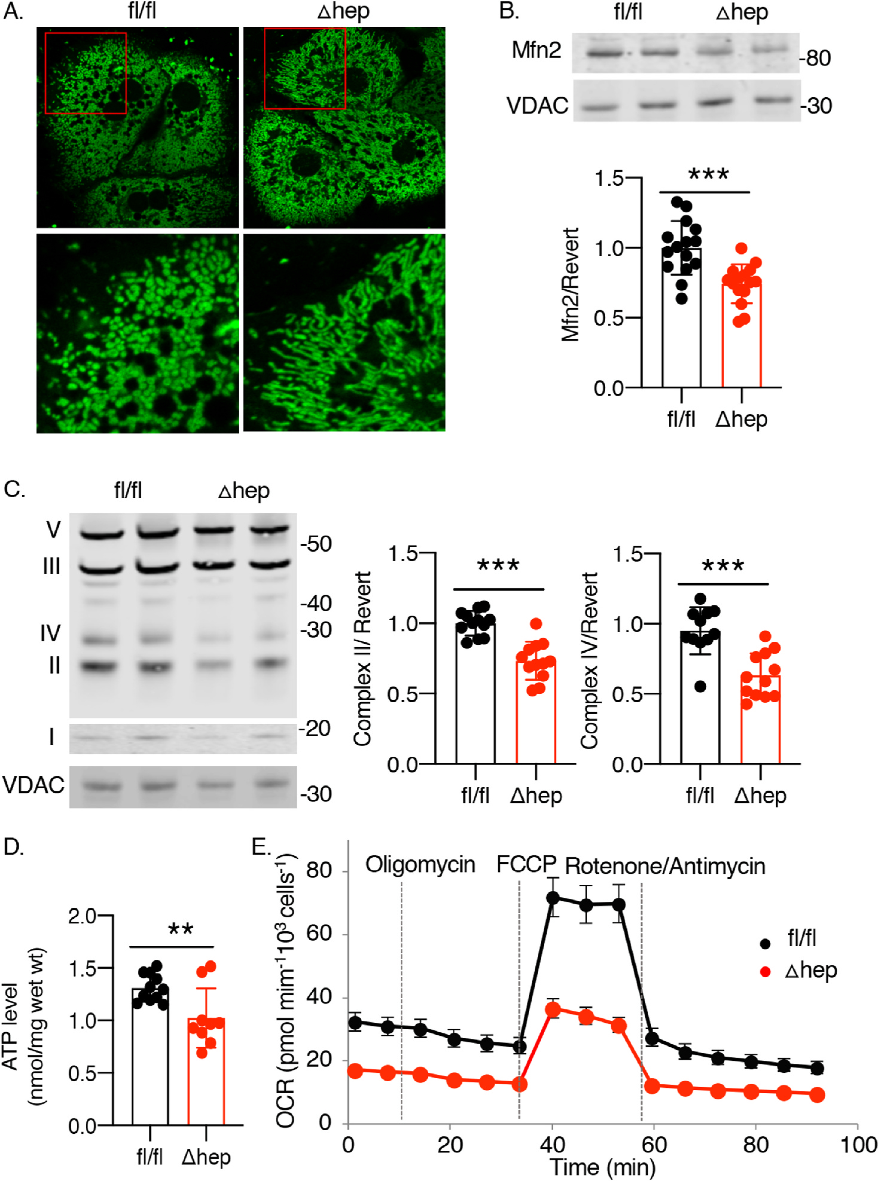
Fig. 2. PCBP1Δhep mice livers exhibit mitochondrial dysfunction.

A. Elongated and hyperfused mitochondria in hepatocytes from PCBP1Δhep livers. Hepatocytes from PCBP1 fl/fl and Δhep mice were isolated and stained with mitochondrial-specific fluorescent dye Mitotracker green and subjected to live-cell confocal imaging. Inset (red) magnified in lower panels. B. Depletion of mitochondrial fusion protein mitofusin 2 in PCBP1Δhep livers. Mfn2 levels measured by quantitative Western blot with VDAC as mitochondrial loading control. C. Depletion of mitochondrial respiratory complexes C II and C IV in PCBP1Δhep livers. Western blot analysis of liver lysates probed with mixed antibodies for respiratory complexes I (NDUFB8), II (SDHB), III (UQCRC2), IV (MTCO1), and V (ATP5A). Quantification of C II and IV shown on right. D. ATP depletion in livers from PCBP1Δhep mice. Data represent mean ± SD. * indicates p < 0.05, **p < 0.01, ***, p < 0.001 as determined by unpaired t-test (B, D) or 2-way ANOVA with Bonferroni multiple comparison test (C). E. Reduced mitochondrial oxygen consumption in hepatocytes from PCBP1Δhep livers. Primary hepatocytes were isolated and mitostress test was performed to evaluate the mitochondrial oxygen consumption. Data from representative experiment are shown, see also Supplemental Fig. 2.
