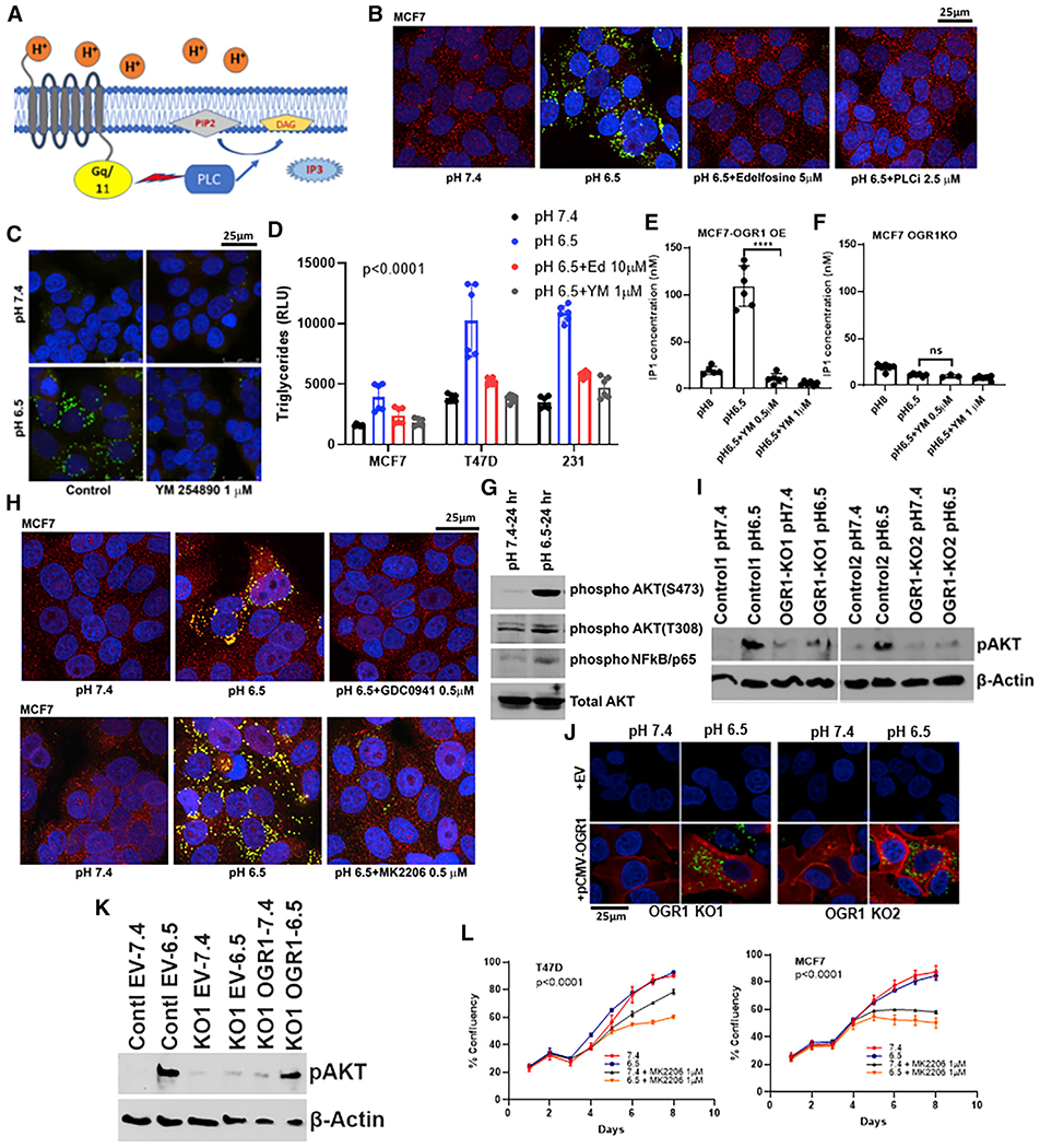
Figure 4. Lipid-droplet formation under low pH is affected by inhibition of downstream signaling of PL-C and PI3Kinase/Akt pathway.

(A) Schematic showing signaling through OGR1 receptor.
(B) LD accumulation is inhibited by pharmacological inhibitors of PL-C, edelfosine (5 μM), or U73122 (2.5 μM) under low pH in MCF7 cells.
(C) MCF7 cells treated with YM254890, an inhibitor of Gq/11-coupled GPCR signaling, affected LD accumulation. PLIN2 (red), Nile Red (green), and DAPI (blue).
(D) Triglyceride levels from cells grown in pH 7.4, 6.5, or 6.5 media along with inhibitors of OGR1 downstream signal mediators PL-C and Gq/11. Acidic-pH-induced increases in TG levels were abrogated by inhibitor treatments. p < 0.0001, one-way ANOVA analysis for each cell line.
(E) Acidic pH induced OGR1-mediated Gq/11 activation as seen by IP1 assay. IP1 levels in OGR1-overexpressing MCF7 cells were higher at pH 6.5 compared with 8 and were inhibited by Gq/11 inhibitor (T test, p < 0.0001).
(F) OGR1-depleted cells did not induce IP1 upon low-pH treatment, and this was not significantly altered by Gq/11 inhibitor. t test, not significant.
(G) Acidic pH induces Akt phosphorylation (residues S473 and T308) and phosphorylation of NFkB, the downstream target of Akt activation, as seen by western blot analyses.
(H) Inhibition of PI3K using GDC0941 (0.5 μM) or Akt inhibition by MK2206 (0.5 μM) resulted in significantly lower levels of LDs under low pH in MCF7 cells.
(I) Acid-induced Akt phosphorylation (S473) was abrogated in OGR1-depleted cells, as seen by western blot analysis.
(J) Ectopic expression of OGR1 rescued acid-induced LD accumulation in OGR1-depleted cells. OGR1-KO cells were transfected with either empty vector control or Myc-tagged OGR1 and subsequently induced with pH 7.4 or 6.5 media (Nile Red, green; Myc, red; DAPI, blue).
(K) Akt phosphorylation (S473) was rescued in OGR1-depleted cells by ectopic expression of OGR1.
(L) Akt inhibition is selectively cytotoxic at acidic pH. Akt inhibitor MK0026 treatment decreased growth rate in low-pH culture conditions compared with neutral pH. p < 0.0001, two-way ANOVA.
