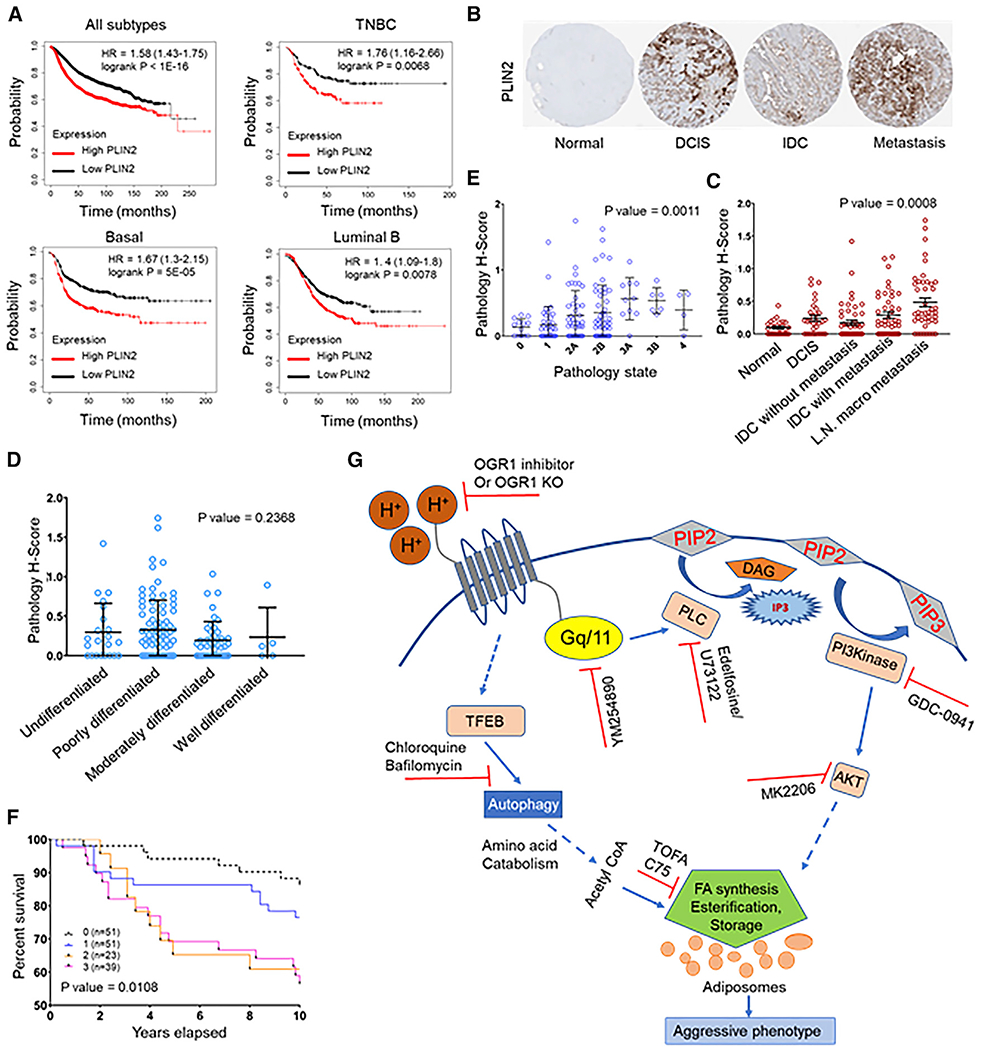
Figure 7. PLIN2 expression is associated with poor patient survival.

(A) Kaplan-Meier survival plot showing significantly low survival of patients with high PLIN2 expression. High expression of PLIN2 is associated with poor prognosis of patients with TNBC, basal, and luminal B subtype of breast tumors (KM plotter).
(B) Representative images from breast cancer progression tissue microarray (TMA) stained for PLIN2.
(C) Increase in PLIN2 expression correlates with aggressive/metastatic tumors (p = 0.0008).
(D) Expression of PLIN2 did not significantly differ between undifferentiated, poorly differentiated, and well-differentiated tumors (p = 0.2368).
(E) PLIN2 levels increases with breast cancer progression, as seen from cores representing different stages (p = 0.0011).
(F) KM plot showing significantly low survival of patients with high PLIN2 expression as quantified from the TMA. p = 0.0108.
(G) Schematics of the mechanism by which acid-induced LD accumulation occurs in breast cancer cells. Acidosis triggers signaling through acid receptors such as OGR1 and induces autophagy supporting de novo lipogenesis and LD accumulation. Acid-induced signal-transduction cascade involves activation of Gq/11, phospholipase C, PI3K, and Akt. Inhibition of these signaling events using pharmacological inhibitors (red symbols) dampened acid-induced LD formation. Acid induction of LDs occurs even in de-lipidated serum, indicating the endogenous origin of lipids. Further, acidosis induced autophagy though the master regulator TFEB. Blocking autophagy and lipogenesis also blocked LD accumulation. Catabolism of ketogenic amino acids produced during autophagic proteolysis contribute significantly to the carbon pool for FA synthesis and storage as LDs. These events appear to be important for survival under acid stress, as the predominant LD coat protein PLIN2 gene expression is associated with poor survival in breast cancer. (Dotted arrows represent events involving multiple steps.)
