Figure 3.
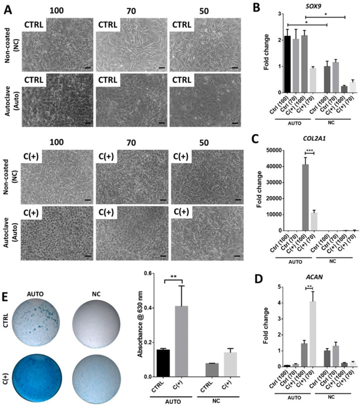
Effect of cell seeding density on chondrogenic differentiation. The hPDCs were seeded on the autoclaved dcECM extract coating (AUTO) and compared to cells on non-coated (NC) tissue culture plastic. The gel coating was air dried at 37 °C for 72 h before seeding. (A) Brightfield images were obtained seven days post-culture; Scale bar = 50 µm. Chondrogenic gene expression of (B) SOX9 (C) COL2A1 and (D) ACAN. The hPDCs were seeded in high density on non-coated surfaces or dcECM coatings under control conditions (CTRL) and chondrogenic C(+) conditions at densities of 100 × 103 (100) 70 × 103 (70) or 50 × 103 (50) cells/well (96). (E) Brightfield microscopy images of Alcian blue (glycosaminoglycan) stained cultures and associated quantification. (n = 3 * p < 0.05, ** p < 0.01, *** p < 0.001).

