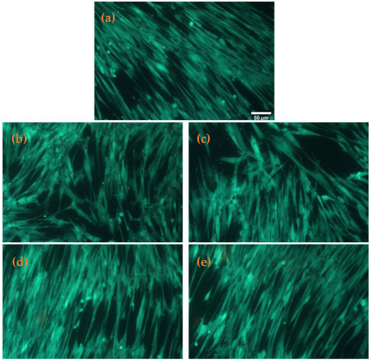
Figure 10.
Fluorescence images of live (green) and dead (red) cells stained with calcein AM and ethidium, respectively, after 24 h incubation of MRC-5 human fibroblasts with (a) control, (b) Fe3O4@STR, (c) Fe3O4@NEO, (d) PLGA-CS-Fe3O4@STR and (e) PLGA-CS-Fe3O4@NEO spheres (scale bar is 50 µm and it is the same for all images).

