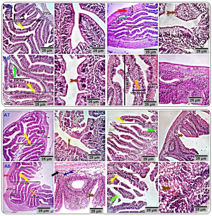
Figure 3.
Stained sections of the anterior and posterior intestines of the respective treatment groups (CUR200, CUR400, CUR600, and CUR800) showed normal histomorphological structures with an increase in the villus height and crypt depth (yellow and green arrows), goblet cell number (brown arrows), and the number of mucosal and submucosal immune cells (black arrows). A moderate number of eosinophilic granular cells are seen in the submucosal tissue of all experimental groups, particularly groups CUR400 and CUR600 (blue arrow). Scale bars = 25 µm. (A5: CUR200, A6: CUR400, A7: CUR600, and A8: CUR800).

