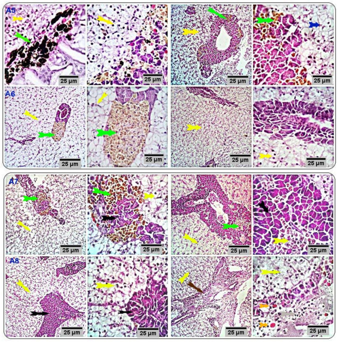
Figure 4.
Stained sections of the liver of the respective treatment groups (CUR200, CUR400, CUR600, and CUR800) showing histomorphological structures comparable to that of the control group (yellow and black arrows) with activated hepatopancreatic acini and normal vascular structures with few surrounding numbers of melano-macrophages. Other sections showed marked infiltration of melano-macrophages, particularly at the hepatic portal/pancreas (green arrows) with occasional vascular dilation, edema, infiltration of eosinophilic granular cells (orange arrows), and pancreatic acinar disorganization and/or degeneration (brown arrow); some of the hepatocytes appeared degenerated (blue arrow). Scale bars = 25 µm. (A5: CUR200, A6: CUR400, A7: CUR600, and A8: CUR800).

