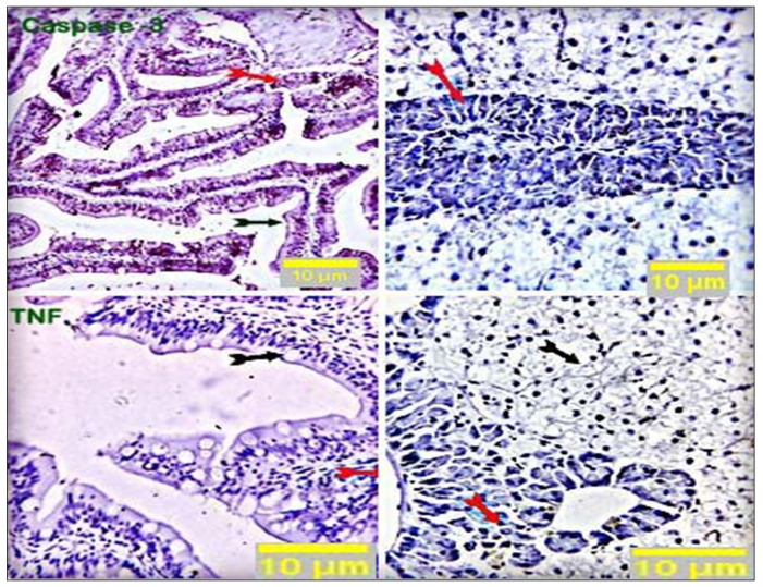
Figure 5.
Stained sections of the intestine and liver of the control group showed negative staining reactions in all the examined parts of the intestinal and hepatic tissue (black and red arrows) against caspase-3 and TNF-α antibodies. Nearly 10% of hepatic portal cells reacted positively to caspase-3. Scale bars = 10 µm.

