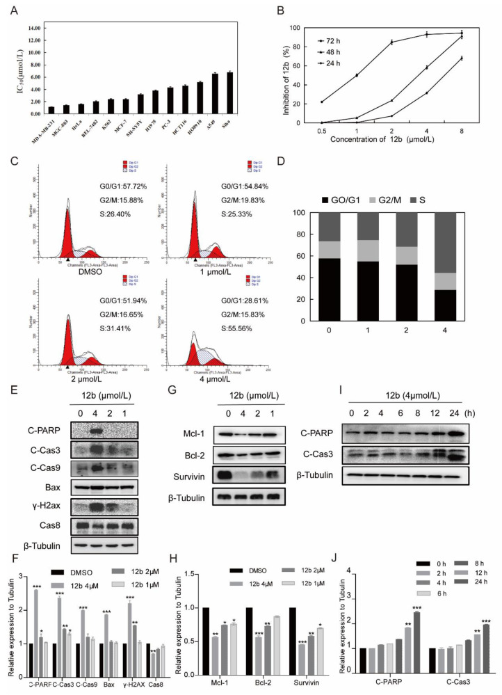
Figure 3.
12b inhibits various cells’ viability and induces MDA-MB-231 cells apoptosis. (A) IC50 values of 12b on various tumor cells. Cancer cells were treated with the indicated concentration of 12b for 72 h. Cell proliferation was determined by using an MTT assay. (B) Proliferation inhibition of 12b on MDA-MB-231 cells. Cells were treated with the indicated concentration of 12b for 24, 48, and 72 h. Cell proliferations were determined by MTT assay. (C) 12b affected cell cycle distribution. Cells were treated with 12b (1–4 μM) for 24 h, then were collected and stained by PI. The DNA content of cells was determined with the MoFlo XDP flow cytometry system. (D) Histograms showed the percentage of cells in G0/G1, G2/M, and S phase after treatment with 12b. (E–J) Effects of 12b on the expression of proteins related to apoptosis. MDA-MB-231 cells were treated with 12b (1, 2, 4 μM) for 24 h or treated with 12b (4 μM) for indicated times. Protein levels were analyzed by Western blotting. The relative band intensities of proteins to the β-Tubulin were analyzed by Image J software. Protein band densities were quantified by normalizing to the DMSO group. Data are presented as means ± SD. All experiments were performed in three replicates. * p < 0.05, ** p < 0.01, *** p < 0.001.

