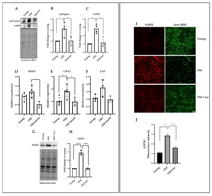
Figure 4.
Taurine treatment attenuates oxidative stress in old mice. (A) Western blot analyses of total lysates obtained from TA muscles were carried out to evaluate the levels of G6PD and Gp91phox. A representative blot is shown. (B,C) Densitometric analysis was performed using a stain-free blot to verify the loading of the samples. (D–F) Real-time PCR analysis of SOD1, GPX1, and CAT expression in TA muscles treated as indicated above. (G) Western blot analyses of total lysates obtained from TA muscles were carried out to evaluate the levels of SOD1. A representative blot is shown. (H) Densitometric analysis was performed using a stain-free blot to verify the loading of the samples. (I) Immunofluorescence analysis of 4-HNE used as a marker of oxidative stress and slow-MHC expression. (J) Quantification of 4-HNE fluorescence intensity. Statistical analysis was performed using one way ANOVA multiple comparison * p < 0.05, *** p < 0.001, **** p < 0.0001, n ≥ 3 mice per group. Data are represented as mean ± SEM. •, ■ and ▲ represent samples from Young, Old, and Old+ taurine groups.

