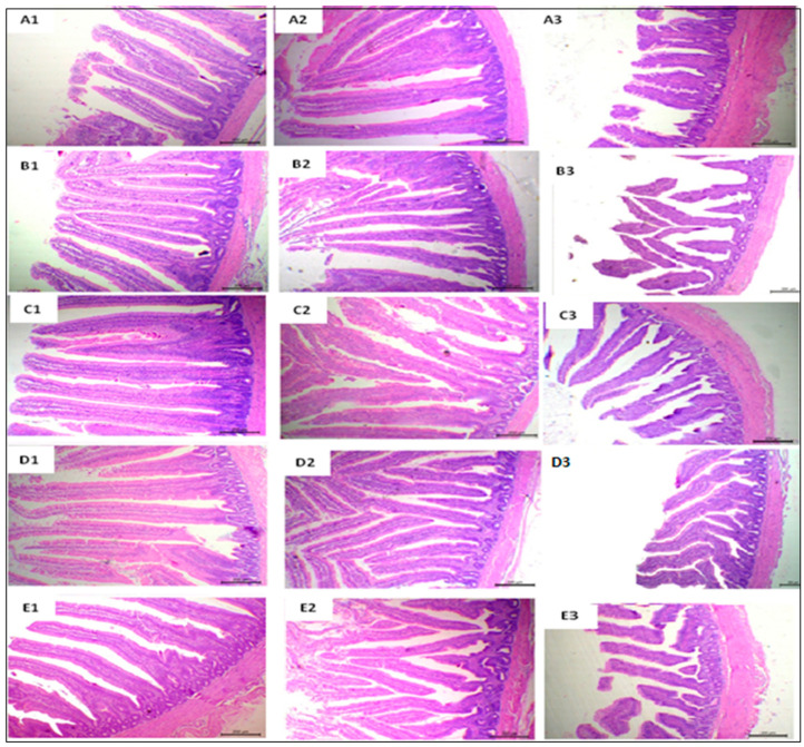
Figure 2.
Representative photomicrograph of H&E-stained small intestine sections of broiler chickens in 40× magnification. Duodenal sections from SPC0 showed normal intestinal villi with a free lumen (A1); those from SPC0.25 group showed a mild increase in villus height (B1); sections from SPC0.5–1 groups revealed markedly thin, tall, and separate villi with mild goblet cell proliferation, increased sizes, and rows of enterocytes with arranged lamina propria (C1,D1,E1). Jejunal segment sections of SPC0 showed free lumina with nearly normal villus structures (A2), while sections from the SPC0.25 group showed a small increase in villi length (B2). Jejunum segments from SPC0.5−1 groups showed closely packed villi with a marked increase in their length and markedly serrated surfaces with goblet cell metaplasia (C2,D2,E2). Ileal segment sections from basal treatment showed tongue-shaped villi with a different height (A3); sections from the SPC0.25 group exhibited normal histology (B3); the sections from the SPC0.5 and SPC0.075 groups showed relatively increased villus height (C3,D3); those from the SPC1 diet group showed normal villi (E3).

