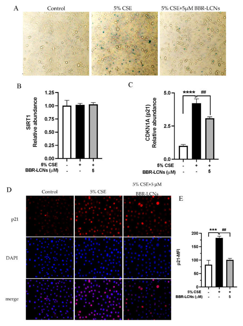
Figure 3.
Effect of BBR-LCNs on 5% CSE-induced senescence of 16HBE. 16HBE cells treated with BBR-LCNs and 5% CSE for 24 h. (A) Cells were stained with b-galactosidase staining kit. Senescence-positive cells are represented with blue-colour positive staining of x-gal. Microscopic images were captured under a 20× magnification. Gene expression of (B) SIRT1 and (C) CDKN1A (p21). **** p < 0.0001 vs. control (without BBR-LCNs and 5% CSE treatment) and ## p < 0.01 vs. 5% CSE. (D) Immunocytochemistry staining of p21-Alexa647; microscopic images were captured at 40× magnification. (E) Mean fluorescence intensity (MFI) of p21. *** p < 0.0001 vs control (without BBR-LCNs and 5% CSE treatment) and ## p < 0.01 vs. 5% CSE.

