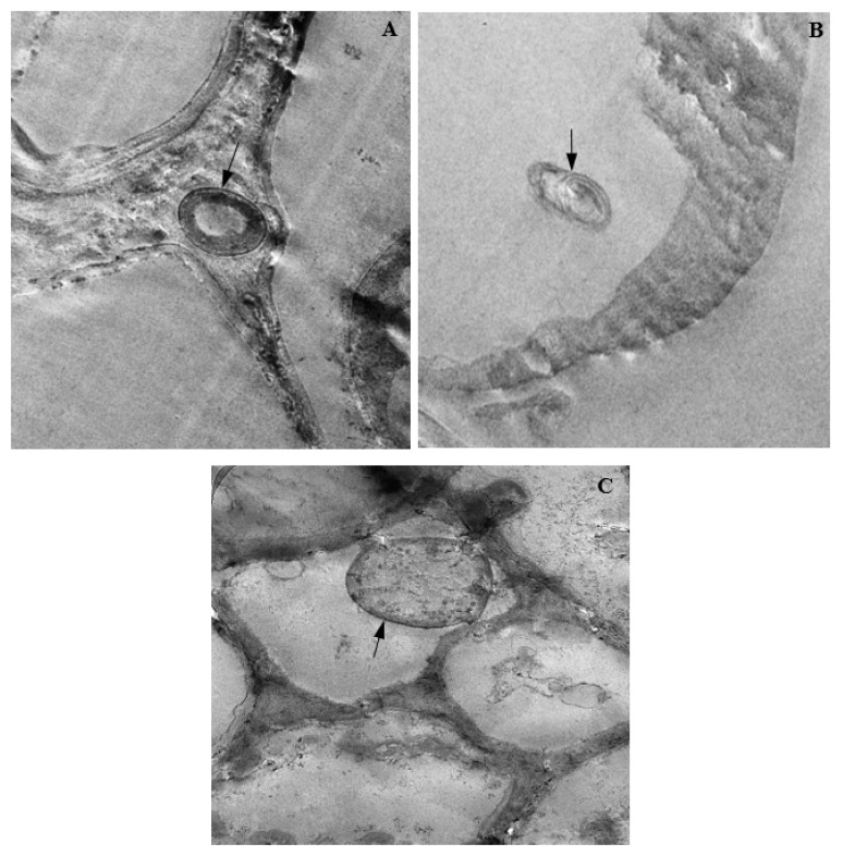
Figure 13.
Transmission electron micrographs showing endophytic colonization of corn tissues by Fusarium verticillioides. The black arrows indicate the presence of hyphae in infected the (A) root intercellular space (Magnification 4000×); (B) stem parenchyma (Magnification 3200×); and (C) leaf mesophyll (Magnification 5000×).

