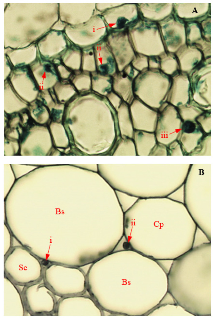
Figure 14.
Light micrographs showing endophytic colonization of corn tissues by Fusarium sacchari (Magnification 400×). Red arrows indicate position of endophyte in infected (A) i: root endodermis; ii: root pericycle; and iii: root pith; (B) intercellular spaces between (B) i: stem sclerenchyma (Sc) and bundle sheath cells (Bs); and ii: stem bundle sheath cells (Bs) and cortical parenchyma (Cp).

