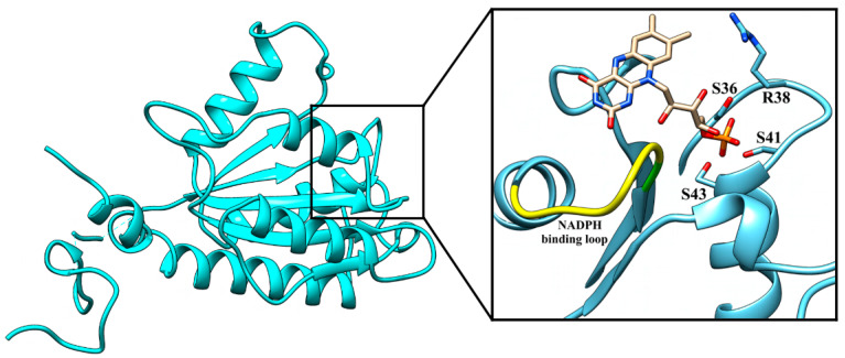
Figure 1.
Overall structure of ArsH from P. denitrificans and the putative active site domain. The image was generated with Chimera 1.13.1 on the basis of the ArsH crystal structure (7PLE). Superposition of FMN from FerB (white) and ArsH in its an active site (zoomed in the square). The 3D coordinate data of FMN were taken from the PDB file 3U7R. The amino acids that probably interact with the phosphate group of the coenzyme and the putative NADPH binding loop are shown.

