Figure 5.
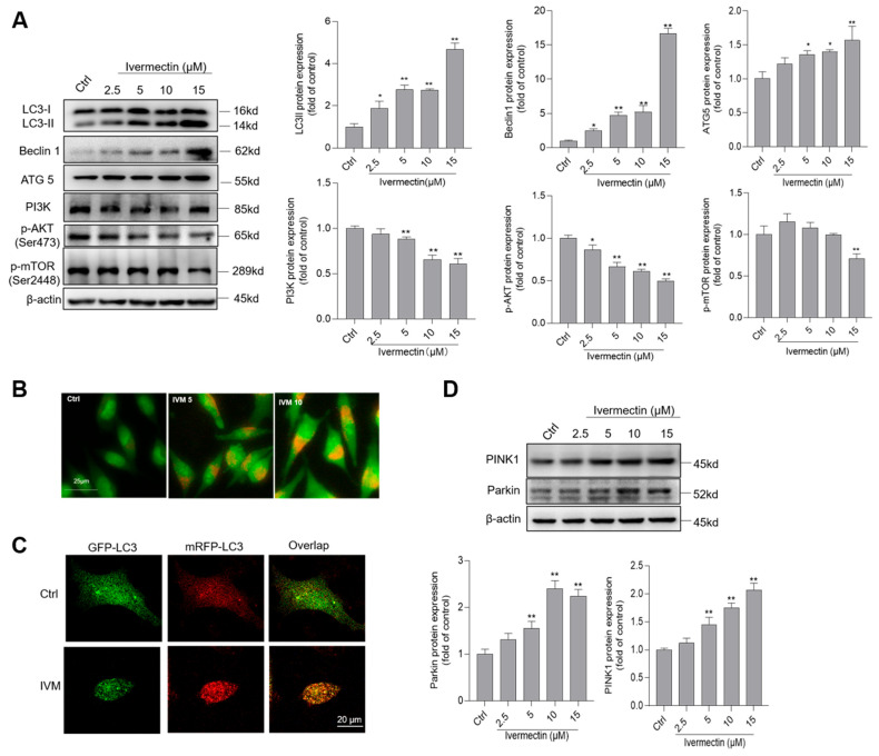
Ivermectin (IVM) induces autophagy and inhibits Akt/mTOR pathways in human SH-SY5Y cells. (A) the expressions of LC3II, Belcin1, ATG5, PI3K, p-Akt (ser 473), and p-mTOR (ser 2448) proteins in IVM (at 2.5, 5, 10, and 15 μM for 24 h)-treated cells were examined by using Western blotting. The representative gel (on the left) and quantitative analysis (on the right) are shown. (B) acidic vesicular organelles were detected using acridine orange (AO) staining. Bar = 25 μm. (C) autophagy flux was monitored by the mRFP-GFP-LC3 plasmid transfection method. Cells were treated with IVM treatment at 5 μM for 24 h, autophagosomes and autolysosome were observed and photographed by a laser scanning confocal microscope. Bar =20 μm. (D) IVM treatment activates mitophagy. The expressions of PINK1 and Parkin proteins were examined. Data shown are represented as the mean ± SD, from three independent experiments (n = 3); compared to the control group, * p < 0.05, ** p < 0.01. CQ 5, chloroquine 5 μM; CQ 10, chloroquine 10 μM.

