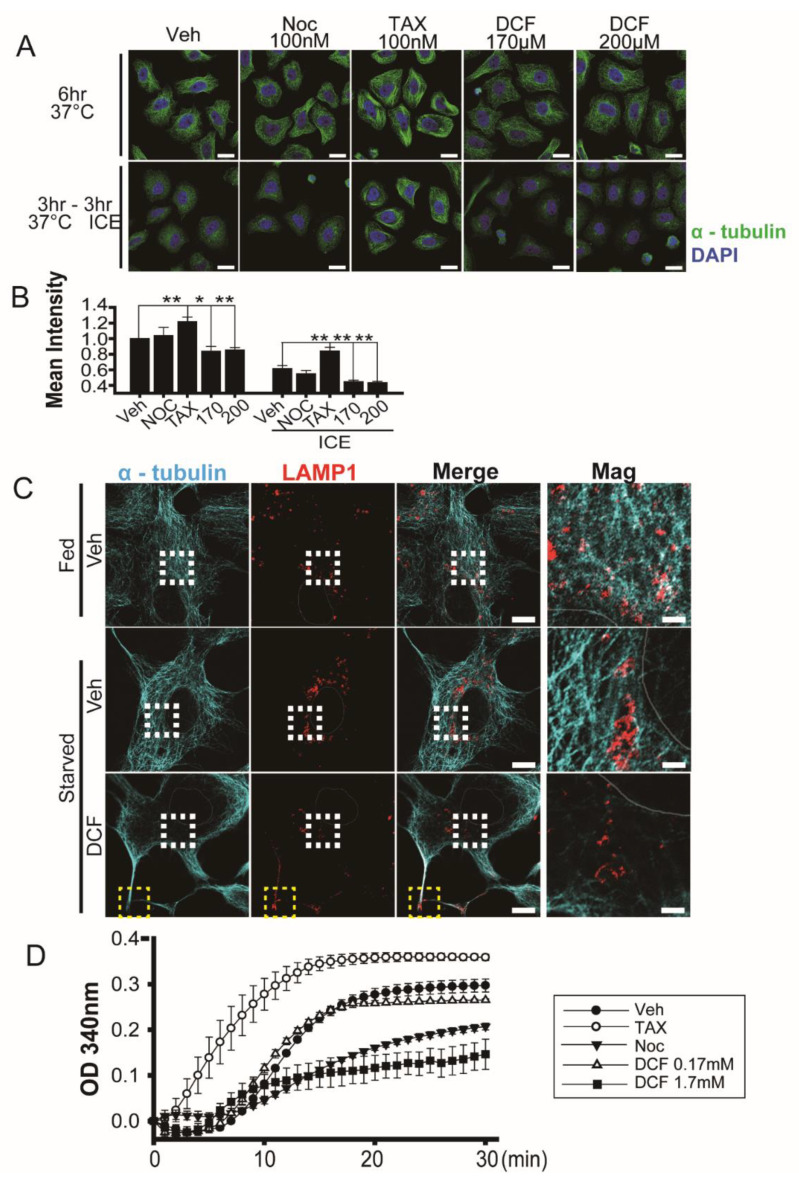
Figure 2.
Diclofenac induces microtubule depolymerization. (A) Confocal microscopy of HeLa cells stained with antibodies to α-tubulin (green) after incubation at 37 °C (upper panel) for 6 h or at 37 °C for 3 h followed by 4 °C for 3 h. Cells were incubated in a medium containing vehicle (0.1% dimethyl sulfoxide [DMSO]), nocodazole (100 nM), taxol (100 nM), or diclofenac (170 µM, 200 µM). Nuclei were stained with 4–6-diamidino-2-phenylindole (DAPI, blue). (B) Quantitative analysis of mean fluorescence intensity of α-tubulin. Data are presented as means ± SD from three independent experiments (n = 52–60 cells). * p < 0.05; ** p < 0.01 (Student’s t-test). (C) Confocal microscopy of HepG2 cells stained with antibodies to α-tubulin (green) and LAMP1 (red) after incubation in a medium containing vehicle (0.1% DMSO) or diclofenac (500 µM) under fed (Dulbecco’s modified eagle medium, 10% fetal bovine serum) conditions or starved (Earle’s balanced salt solution) conditions for 8 h. Nuclei were stained with DAPI (blue). Representative images are shown. (n = 14–18 cell). Areas enclosed by the white boxes are shown at higher magnification. Yellow boxes indicate the edges of the plasma membrane. Three independent experiments were performed. Scale bar, 10 µm; scale bar in magnification; 2 µm. (D) In vitro tubulin polymerization. Polymerization activity was monitored in the presence of DMSO (0.01%, vehicle), taxol (10 μM), nocodazole (10 μM), or diclofenac (0.17 mM and 1.7 mM) for 30 min at 37 °C as the increase in A340 nm. Two independent experiments were performed.

