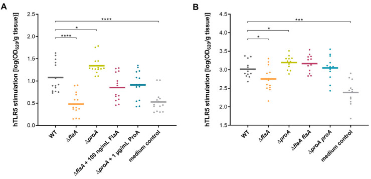
Figure 4.
TLR5 activation in HEK-Blue™ cells by supernatants of infected lung tissue explants (HLTEs). Explanted human lung tissue specimens were infected with the L. pneumophila Corby WT, a proA-negative mutant ΔproA and a flaA-negative mutant ΔflaA. Loss of ProA or FlaA was reconstituted either by addition of purified proteins (1 µg/mL ProA and 100 ng/mL monomeric FlaA) (A) or use of the specific complementation strains ΔproA proA and ΔflaA flaA (B). Untreated tissue samples served as a negative control. Infected tissue pieces were weighed, and supernatants were isolated and incubated with HEK-Blue™ hTLR5 cells for 16 h at 37 °C and 5% CO2. Activation of the TLR5-NF-κB pathway was measured by SEAP activity at OD620 and is shown in scatter plots with means. Compared to the L. pneumophila WT, a HEK-Blue™ Detection assay revealed significantly reduced TLR5 activation by ΔflaA-infected tissue supernatant and elevated stimulation with the ΔproA mutant strain. These effects were apparent after 2 h (A) and 24 h (B) post infection and were successfully restored by complementation with respective proteins or gene sequences. Significance of at least twelve replicates was evaluated by repeated measurement one-way ANOVA with Dunnett’s post hoc test for simple effect analysis (* p ≤ 0.05, *** p ≤ 0.001, **** p ≤ 0.0001).

