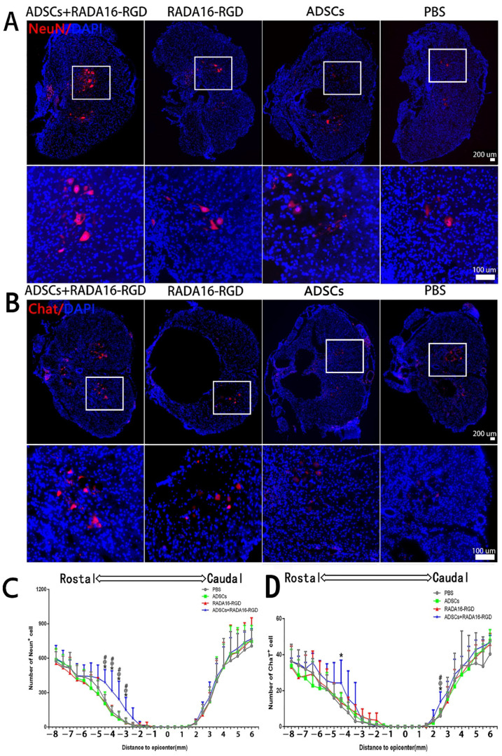
Figure 4.
ADSCs+RADA16-RGD transplantation contributes to neuronal survival in rats with spinal cord injury. (A) NeuN antibody staining (red) at −4.5 mm cephalic to the injury at 8 weeks after PBS, ADSCs, RADA16-RGD, and ADSCs+RADA16-RGD transplantation. (B) ChAT antibody staining (red) at −4.5 mm cephalic to the injury at 8 weeks after ADSCs+RADA16-RGD transplantation. The panels below (A,B) show high magnification of the boxed regions in the abovementioned panels. Scale bar = 200 µm, high magnification regions scale bar = 200 µm. (C) Quantitative analysis of NeuN+ cells transected at different distances from the injury center. The number of NeuN+ neurons on the cephalic side of the injury center was significantly higher in the ADSCs+RADA16-RGD group than in the PBS, ADSCs, and RADA16-RGD groups (ADSCs+RADA16-RGD vs. PBS: p < 0.05, ADSCs+RADA16-RGD vs. ADSCs: p < 0.05, ADSCs+RADA16-RGD vs. RADA16-RGD: p < 0.05, at −3 mm, −3.5 mm, −4 mm, and −4.5 mm). (D) Quantitative analysis of ChAT+ cells transected at different distances from the injury center. The number of ChAT+ neurons was greater in the ADSCs+RADA16-RGD group than in the PBS group at −4.5 mm on the cephalic side of the injury center (p < 0.05). Similarly, the number of ChAT+ neurons was greater in the ADSCs+RADA16-RGD group than in the PBS, ADSCs, and RADA16-RGD groups at 3 mm (ADSCs+RADA16-RGD vs. PBS: p < 0.05, ADSCs+RADA16-RGD vs. ADSCs: p < 0.05, ADSCs+RADA16-RGD vs. RADA16-RGD: p < 0.05) on the caudal side of the injury center. *: ADSCs+RADA16-RGD vs. PBS, @: ADSCs+RADA16-RGD vs. ADSCs, # ADSCs+RADA16-RGD vs. RADA16-RGD. * p < 0.05, @ p < 0.05, # p < 0.05. Data are presented as the mean ± SEM (NeuN n = 6 per group, ChAT n = 6 per group). ADSCs: adipose stem cells; DAPI: 4′6-diamidino-2-phenylindole; NeuN: neuronal nuclei; ChAT: choline acetyltransferase. NeuN (red): Alexa Fluor 647, ChAT (red): Alexa Fluor 647.

