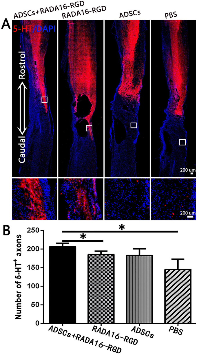
Figure 7.
ADSCs+RADA16-RGD transplantation increases the number of 5-HT+ nerve fibers. (A) At 8 weeks of transplantation of ADSCs+RADA16-RGD, 5-HT antibody staining (red) was performed via passing through the central sagittal section of the spinal cord injury. The panels below (A) show high magnification of boxed regions in top panels. (B) Quantitative analysis of 5-HT+ nerve fibers at the injury center. The ADSCs+RADA16-RGD group was significantly higher than in the PBS and RADA16-RGD groups (ADSCs+RADA16-RGD vs. PBS or RADA16-RGD: p < 0.05). No 5-HT+ nerve fibers could be found in the three groups below the caudal edge of the injury center. * p < 0.05. Data are presented as mean ± SEM (n = 3 per group). Scale bar = 200 µm. ADSCs: adipose stem cells; 5-HT:5-hydroxy tryptamine; DAPI: 4′6-diamidino-2-phenylindole. 5-HT (red): Alexa Fluor 647.

