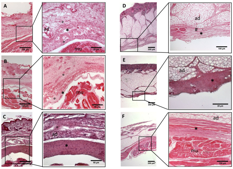
Figure 4.
Histological images (hematoxilyn-eosin) of lower limb, leg: crural fascia development. (A): 24 weeks; (B): 27 weeks; (C): 29 weeks; (D): 36 weeks; (E): 38 weeks; (F): 40 weeks. (*): deep fascia; (°): mesenchymal cells; (#): areolar tissue; (mu): muscle; (ad): adipose tissue. The first part of gestation is distinguished (A–C) by an irregular connective tissue without clear organization in layers, whilst the second part of the gestation (D–F) showed up with a denser connective tissue, and was better organized in the fascial layers.

