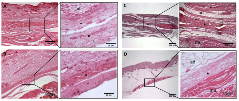
Figure 5.
Histological images (hematoxilyn-eosin) of lower limb: extensor retinaculum development. (A): 24 weeks; (B): 29 weeks; (C): 38 weeks; (D): 40 weeks. (*): deep fascia; (°): mesenchymal cells; (#): areolar tissue; (mu): muscle; (ad): adipose tissue. The first part of gestation is distinguished (A,B) by an irregular connective tissue, whilst the second part of the gestation (C,D) showed up with a denser connective tissue, which was better organized in the fascial layers.

