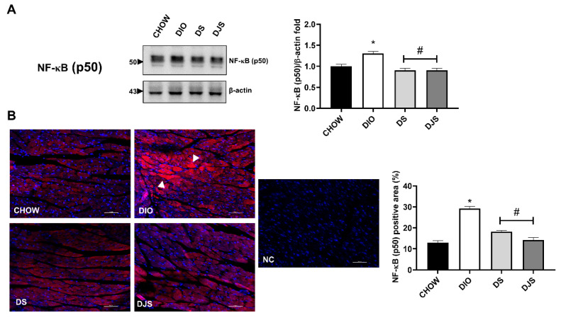
Figure 4.
Measurement of nuclear factor kappa-light-chain-enhancer of activated B cells subunit p50 (NF-κB p50). (A) Cardiac lysates from rats were immunoblotted using specific anti NF-κB (p50). Graph shows the ratio of densitometric analysis of bands and β-actin expression used to normalize the data, taking CHOW rats as a reference group; (B) Confocal image of representative immunofluorescent staining for NF-κB (p50) in the heart and quantification expressed as percentage (%) of NF-κB (p50) positive area. Magnification 10× zoom 3. Scale bar 10 µm. NC, Negative control. Arrowheads indicate the more immunoreactive cardiomyocytes. CHOW rats (n = 8), fed with standard diet; DIO rats (n = 9), fed with high-fat diet; DS (n = 12), DIO rats supplemented with tart cherry seeds; DJS (n = 9), DS rats supplemented with tart cherry juice. Data are mean ± SEM. * p < 0.05 vs. CHOW rats; # p < 0.05 vs. DIO rats.

