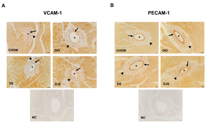
Figure 6.
Immunohistochemical analysis of inflammatory adhesion molecules. Representative pictures of heart sections processed for the immunohistochemistry of vascular cell adhesion molecule-1 (VCAM-1) (A) and platelet endothelial cell adhesion molecule-1 (PECAM-1) (B). The immunoreaction is located in the endothelium (arrows) while the tunica media (arrowheads) is negative. The lumen of vessels is marked with an asterisk (*). Magnification 40×. Scale bar 25 µm. NC, Negative control. CHOW rats (n = 8), fed with standard diet; DIO rats (n = 9), fed with high-fat diet; DS (n = 12), DIO rats supplemented with tart cherry seeds; DJS (n = 9), DS rats supplemented with tart cherry juice.

