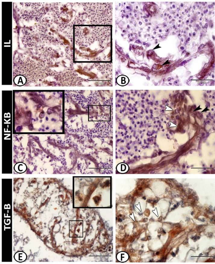
Figure 6.
Immunohistochemistry of IL-1β, NF-κB, and TGF-β in the spleen. (A,B) IL-1β showed immunoreactivity in monocytes and macrophages around the ellipsoids (arrowheads). (C,D) NF-κB was expressed in the macrophages (boxed areas, white arrowheads) and epithelial reticular cells (black arrowheads). (E,F) TGF-β was expressed in the macrophages (boxed areas) and reticular cells (arrowheads).

