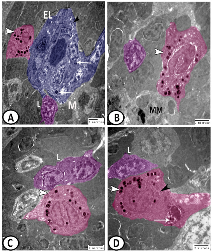
Figure 10.
Digital colored TEM of the leukocytes in the spleen. (A) The ellipsoids (blue, E) were lined with a simple cuboidal epithelium and contained lysosomes (arrows), and vacuoles (black arrowhead). Note the surrounding macrophage (M), lymphocytes (L, violet), and eosinophils (pink, white arrowhead). (B) Eosinophils (pink, arrowhead) displayed horseshoe-shaped nuclei and rounded electron-dense granules and vesicles. Note the presence of lymphocytes (L, violet) and melanomacrophages (MM). (C) The cytoplasm of basophil (pink, arrowhead) was packed with electron-dense granules. Note the associated lymphocytes (L, violet). (D) Neutrophils (pink) with a segmented nucleus, and many electron-dense granules (white arrowhead), rER (black arrowhead), and phagocytosed materials (arrow). Note the presence of lymphocytes (L, violet).

