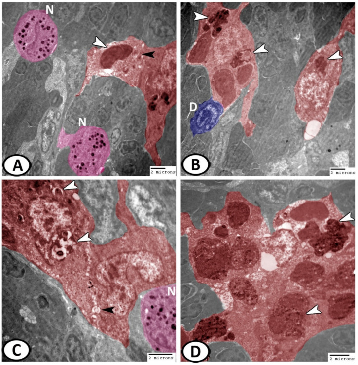
Figure 11.
Digital colored TEM of the macrophages in the spleen. (A,B) Macrophages (red) displayed long pseudopodia and their cytoplasm contained many lysosomes (black arrowheads) and other phagocytosed materials (white arrowheads). They were usually associated with dendritic cells (blue, D) and neutrophils (pink, N). (C) The active phagocytosing macrophages (red) were characterized by the kidney-shaped nucleus, vacuoles (black arrowheads), and phagocytosed materials (white arrowheads). Note the presence of neutrophils (pink, N). (D) They were merged into spherical structures known as melanomacrophage centers (MMCs, red) with vesicles containing pigments (arrowheads).

