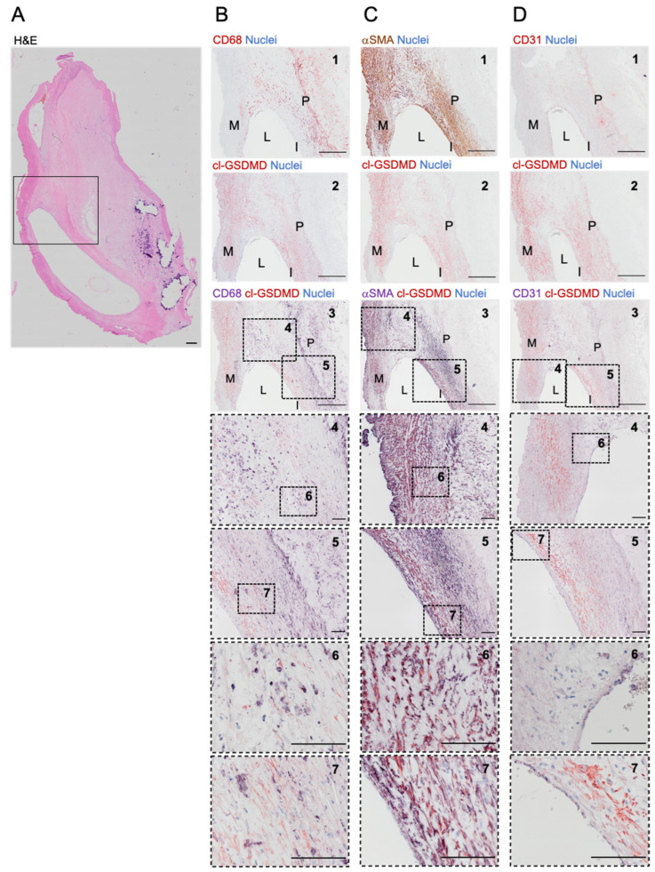
Figure 3.
Cleaved GSDMD is expressed in human carotid plaques. (A) Overview image of a section from a human carotid artery lesion stained with hematoxylin/eosin. The boxed area corresponds with the region shown in (B–D). (B–D) Immunohistochemical staining of cleaved (cl)-GSDMD combined with (B) CD68, (C) α-smooth muscle actin (αSMA) or (D) CD31. From top to bottom: 1. Image of section stained for (B) CD68 (red), (C) αSMA (brown), or (D) CD31 (red). 2. Image of section stained for cl-GSDMD (red). 3. Image of double stained section for cl-GSDMD (red) combined with (B) CD68, (C) αSMA, or (D) CD31 (purple). 4–7. Magnifications of dotted frames in images 3–5. Scale bar = 500 μm (A,B–D: 1–3), 100 μm (B–D: 4–7). Representative images are shown. M = media, L = lumen, I = intima, P = plaque.

