Figure 5.
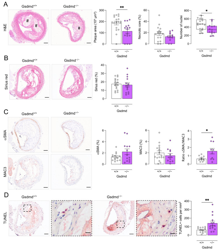
Plaques in the brachiocephalic artery from ApoE−/− Gsdmd−/− mice are smaller but show increased apoptosis. ApoE−/− Gsdmd−/− and ApoE−/− Gsdmd+/+ mice were fed a WD for 16 weeks. Sections of the brachiocephalic artery were stained with (A) hematoxylin/eosin to quantify plaque size, necrotic cores (# hash signs), and cell infiltration; (B) Sirius red to measure total collagen content; (C) anti-MAC3 and anti-α-smooth muscle actin (αSMA) to determine macrophage and smooth muscle cell content, respectively, and to calculate the ratio of αSMA/MAC3 immunoreactivity; (D) TUNEL to count apoptotic cells (dotted boxes are magnified, scale bar = 20 μm). * p < 0.05, ** p < 0.01 (independent samples t-test, boxplot: Mann–Whitney test, n = 10–18 mice per group). Scale bar = 100 μm. Representative images are shown.

