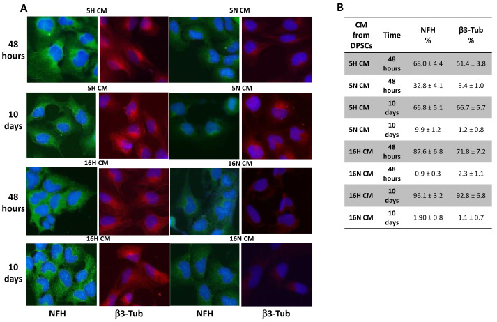
Figure 4.
Immunofluorescence and flow cytometry analysis of SH-SY5Y. (A) SH-SY5Y exposed to DPSC-CM (5H, 16H) were stained for neuronal markers NFH and β3-Tubulin that were highly expressed in all groups of SH-SY5Y treated with DPSCs’ hypoxic CM (5H, 16H) in comparison with groups of cells treated with DPSCs’ normoxic CM (5N, 16N). (B) Flow cytometry analysis of NFH and β3-Tubulin expression in SH-SY5Y-treated groups or untreated cells. Scale bar 20 µm.

