Figure 3.
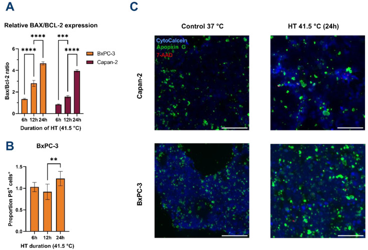
Hyperthermia induces apoptosis in PDAC cells. (A) BAX/BCL-2 expression ratio significantly increases over HT duration for both cell lines. Values were normalized for GAPDH expression and calculated relative to controls at 37 °C. (B) Expression of phosphatidylserine (PS) on the outer membrane is significantly increased after 24 h of HT only for BxPC-3. Data are normalized to controls at 37 °C. Data for Capan-2 are shown in Supplementary Figure S1. No significant differences in PS positivity were observed for Capan-2. (C) Representative fluorescence microscopy imaging of BxPC-3 and Capan-2 cells at 37 °C (control) and after 24 h at 41.5 °C. Viable cells are stained blue, apoptotic cells are stained green (PS), and necrotic nuclei are stained red. Images were acquired at 4× magnification. Scale bars represent 200 µm. All error bars represent 95% CI. Statistical significance ** p < 0.001; *** p < 0.0001; **** p < 0.00001.

