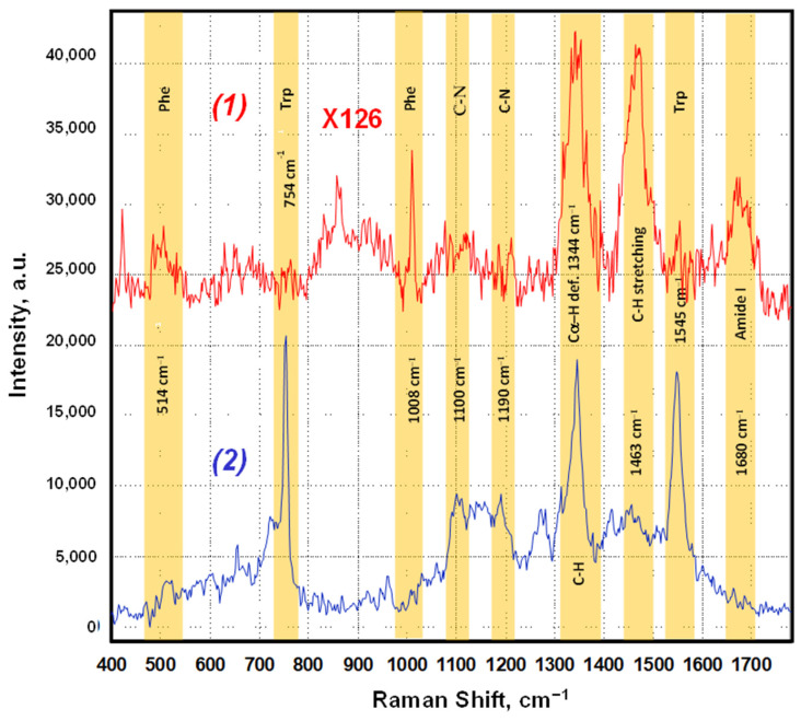
Figure 7.
SERS spectral features of the SARS-CoV-2 RBD with reduced disulfide bonds. (1) An unenhanced Raman spectrum obtained from the native SARS-CoV-2 RBD from a dried drop on a glass slide, about 1.3 pg of RBD. The scaling factor for the integrated intensity is 126. (2) A SERS spectrum obtained from about 1 fg of the sample of SARS-CoV-2 RBD with reactive thiol groups (reduced disulfide bonds) on the silver surface of the SERS-active substrate demonstrating the dominance of the Trp amino-acid residue signals.

