Figure 1.
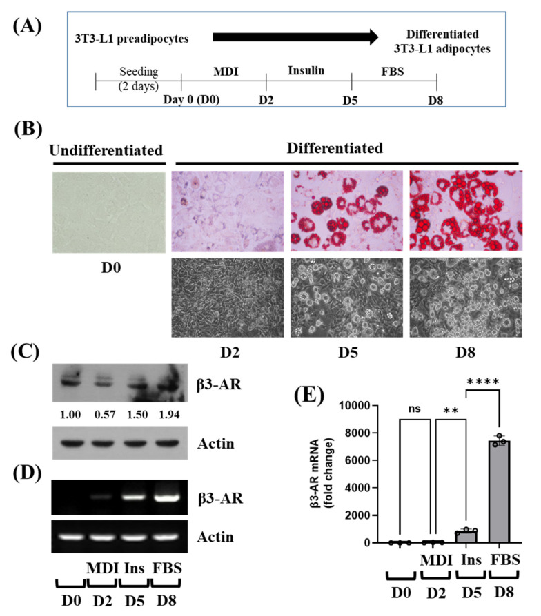
Lipid accumulation and expression of β3-AR at the protein and mRNA levels during 3T3-L1 preadipocyte differentiation. (A) The scheme for 3T3-L1 preadipocyte differentiation. (B) Measurement of lipid droplets (LDs) accumulation on day 0 (D0), D2, D5, and D8 of 3T3-L1 preadipocyte differentiation by Oil Red O staining (upper panels) and by phase-contrast image (lower panels). (C) 3T3-L1 preadipocytes were differentiated with an induction medium containing MDI, insulin, and FBS, and harvested at D0, D2, D5, and D8, respectively. At each time point, whole-cell lysates were prepared and analyzed by immunoblot analysis with respective antibodies. Relative intensities were measured by ImageJ software (version 1.8.0; National Institutes of Health). (D–E) 3T3-L1 preadipocytes were differentiated with an induction medium containing MDI, insulin, and FBS, and harvested at D0, D2, D5, and D8, respectively. At each time point, total cellular RNA was extracted and analyzed by RT-PCR (D) or real-time qPCR (E) with respective primers. In (E), Error bars are indicated as mean ± SD. ** p < 0.01; **** p < 0.0001; ns, not significant, calculated by one-way ANOVA with Sidak’s multiple comparison test.

