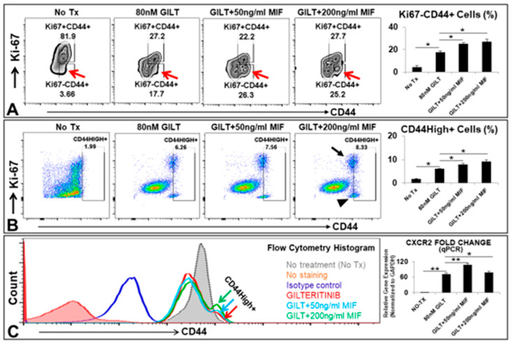
Figure 3.
MIF promoted the survival of a group of CD44High+ cells after TKI treatment in vitro. (A) Representative FC plots of Ki-67 and CD44 expressions in MV4-11 experimental groups after 5 days’ sequential coculture of 80 nM GILT and appropriate doses of MIF in vitro; Red arrow indicates viable Ki-67-CD44+ cells; Right bar chart: Cumulative FC percentage data of viable Ki-67-CD44+ cells; (B) Representative FC plots of Ki-67 and CD44 expressions in MV4-11 experimental groups after 5 days’ simultaneous coculture of 80 nM GILT and appropriate doses of MIF in vitro; Black arrow or arrowhead indicates Ki-67+CD44+ or Ki-67-CD44+ cell population respectively; Right bar chart: Cumulative FC percentage data of viable CD44High+ cells; (C) Representative FC histogram plot of CD44 expression in MV4-11 experimental groups after 5 days’ simultaneous coculture in vitro; Color arrows indicate groups treated with 80 nM GILT alone or its combination with different doses of MIF, showing GILT-treated groups with the supplementation of MIF had a group of CD44High+ cells when compared to the non-treatment control or GILT alone; Right bar chart: qPCR Data show the change of mRNA expression of CXCR2 gene in MV4-11 cells at different doses of MIF combined with 80 nM GILT in vitro; * p < 0.05, ** p < 0.01.

