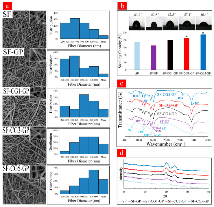
Figure 1.
(a) SEM images and the fiber diameter distribution of all electrospun mats. (b) Wettability of different electrospun mats in terms of swelling capacity and contact angle. (* shows significant differences between each group and SF at p < 0.05). (c) FTIR spectra of all nanofibrous scaffolds. (d) XRD patterns of different samples.

